- Have any questions?
- +86-574-87873760
- +86-15968495319
- admax@kingstic.com

CBM-221C Eye model and orbital model

CBM-221B Half Eye Model(right eye)
29/11/2016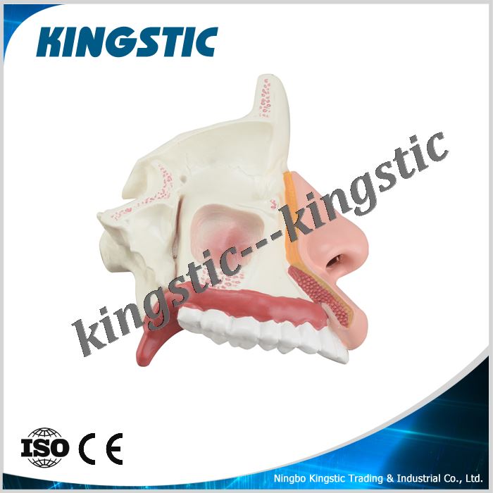
CBM-231A The nasal cavity anatomy model
29/11/2016


Eye model and orbital model
This model shows eye and orbital surrounding structure. The position is under frontal bone, zygomatic above. the 6 muscles can freely change the sight direction, Dissectible into 10 parts, 3 times enlarged. From anterior superficial view, it can help people learn eye muscles, eye nerve, cornea and pupil. From back view, it can help people to learn internal structure of eye including retinal central artery. Environmental PVC with white base.
Reviews
There are no reviews yet.